Accelerate to discover

Back to filter

Related topics

Automatic, Real Time Acquisition of Bioluminescent Kinetic Curves

Spectral Instruments Imaging

Jun 27, 2024

Watch this pre-recorded webinar with Dr. Andrew Van Praagh to learn how our new Aura software feature —Kinetics—...

Theranostics: From Mice to Men and Back

MOLECUBES

Jun 25, 2024

Recorded webinar
Presenters: Prof. Dr. Ken Herrmann and Prof. Dr. Katharina Lückerath – Moderator: Hannah Notebaert

Orion 2024 AACR poster: 17-plex single-step stain and imaging of cell Lung Carcinoma

RareCyte

Jun 21, 2024

RareCyte Orion is a benchtop, high resolution, whole slide multimodal imaging instrument. A combination of quantitative...

Hypoxia in the Tumor Immune Microenvironment (TIME)

Bruker Biospin

Jun 6, 2024

Thursday, 11 July 2024, 16:00 CET | 10:00 EST
Zaver M. Bhujwalla, PhD...

Jul 3, 2024

X-RAD 320 for irradiation therapy during quantifying study for in vivo collagen reorganization

Precision X-Ray

Jun 5, 2024

Quantifying in vivo collagen reorganization during immunotherapy in murine melanoma with second harmonic generation...

Use of MRI and microCT to evaluate gene therapy for the treatment of discogenic back pain

Bruker Biospin

Jun 4, 2024

MRI images were obtained using the 9.4T Bruker BioSpec system, equipped with 40 mm 1H quadrature volume resonator, and...

Exosome-Mediated Delivery of Small Molecules, RNA & DNA for Development of Novel Cancer Therapeutics

Spectral Instruments Imaging

Jun 3, 2024

Disha Moholkar of University of Louisville's Gupta Lab
Tuesday, June 11, 2024, 6:30 PM
...

Jul 3, 2024

Emulate in vivo conditions – introduce shear flow to your experiments with BioFlux system

Cell Microsystems

May 27, 2024

Most research is still conducted in vitro without the presence of flow. We use the BioFlux System to give you the...

Show all topics (10)

CQ1 webinar : optimizing high-content imaging of 3D models for drug discovery

Jan 7, 2021

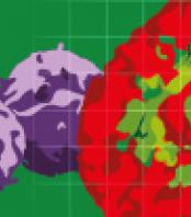
High content imaging and analysis (HCA) of 3D spheroid models can provide valuable information to help researchers untangle disease pathophysiology and assess novel therapies more effectively. Making the move from simple monolayer 2D cell models to dense 3D spheroids in HCI applications, however, requires 3D-optimized protocols, instrumentation, and resources. In this webinar, we will discuss considerations for high content imaging and analysis of 3D spheroid disease models for drug discovery, share lessons we learned while in setting up and conducting proof-of-concept studies designed to test the full potential for high resolution image-based analysis of 3D spheroid models, and provide a working checklist for researchers and core services groups planning to exploit these technologies in their work.

Learning Objectives:

  • High Content assat development- considerations when transitioning from 2D cell models to 3D multicellular models including examples of the rich, physiologically-relevant data that can be extracted from multicellular 3D models
  • 3D image acquisition- high content instrument hardware/software and acquisition settings for efficient acquisition of high quality image stacks
  • 3D image analysis- visualization and image analysis toolsets for 3D imaging, including the application of Deep Learning methods

Arvonn Tully, Senior Software Application Specialist, Yokogawa Life Innovations
JAN 14, 2021 17:00 (CET)

Register now!
Application
Webinar

Related technologies: Live cell imaging

Get more info

Riccardo Pasculli

Head of application support

Riccardo

Pasculli

+420 731 127 717

Send Message

Brand profile

Yokogawa

Yokogawa is a leading provider of confocal quantitative live cell imaging platforms. Combining superior technology with engineering services, project management, and maintenance, Yokogawa delivers field proven operational efficiency, safety, quality, and reliability. 

Related products

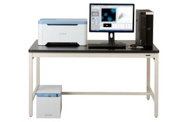
Confocal quantitative image cytometer CQ1 offers a new approach to image quantification

show detail

We supply and support Life-Science Technologies in the territory of Central and Eastern Europe.

Czech Republic

Slovakia

Hungary

Poland

Croatia

Slovenia

Serbia

Romania

Bulgaria

Latvia

Lithuania

Estonia

Russia

Ukraine

Belarus

Turkey