More info at:
s-sharp.com
S-Sharp
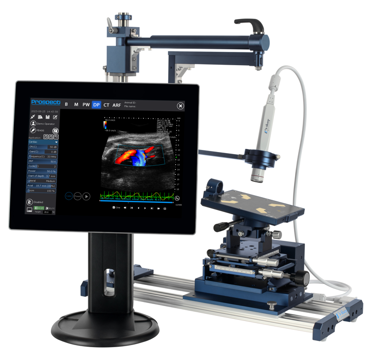
The high-frequency ultrasound instrument
The S-Sharp high-frequency ultrasound instrument enables the researcher to obtain in vivo anatomical, functional, physiological and molecular data simultaneously and in real-time. The system is easy to use, non-invasive and fast, providing extremely high throughput when needed.
Ultrasound imaging is a well-established and validated technology that has been used clinically for many decades to visualize internal organs, soft tissues, tendons, joints and vasculature. S-Sharp has perfected the use of ultrasound in pre-clinical small animal research by creating high-frequency transducers that offers superior high resolution.
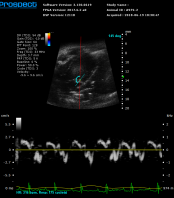
Although B-Mode, which displays a two dimensional cross-section of tissue, is the most common imaging mode with ultrasound, other image types can also be produced: blood flow, localization and direction; vascularity; tissue motion over time; presence of molecules and biomarkers; anatomy and size of a 3D region; tissue stiffness and cardiac strain.


