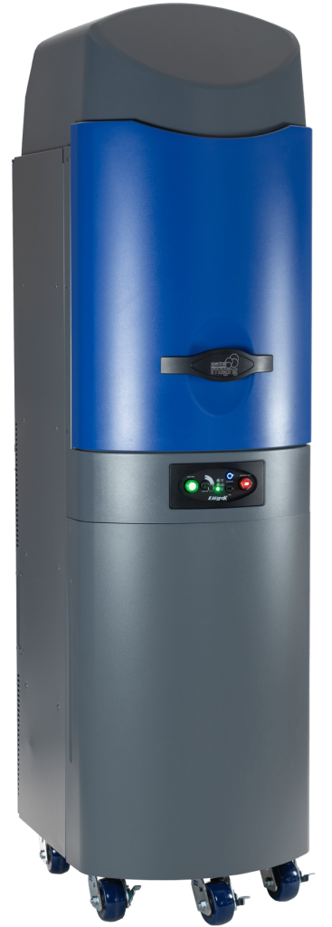
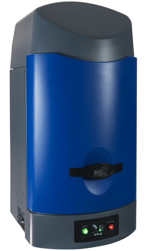
Spectral Instruments Imaging manufactures instruments for preclinical optical (bioluminescent, fluorescent) and X-ray imaging.
Accelerate to discover
Related topics

Optimizing Substrate Dosing for ReliableBioluminescence Imaging (BLI)
Bioluminescence Imaging (BLI) is a powerful tool for monitoring biological processes in vivo, but
achieving consistent and reliable results depends on controlling key experimental variables. One
critical factor is the administration of the luciferase substrate, typically D-luciferin. Variability in
dosing can lead to inconsistent signal intensities, complicating data interpretation. This tech note
outlines best practices for optimizing substrate dosing to enhance reproducibility and data quality.
The Importance of Consistent Dosing
Luciferase-based bioluminescence relies on the enzymatic oxidation of D-luciferin, producing light
that can be detected by your Spectral Instruments Imaging optical imaging system. Uneven
substrate dosing can result in variability in:
ï Signal intensity across subjects.
ï Temporal dynamics of the luminescent signal.
ï Overall data comparability between experiments.
By standardizing dosing, you minimize variability and ensure that observed differences in signal
reflect biological phenomena rather than experimental inconsistencies.
Best Practices for Substrate Dosing
To achieve consistent luciferase substrate administration, consider the following guidelines:
Dose Based on Weight:
Administer D-luciferin based on the animal’s body weight (e.g., 150 mg/kg). This ensures each
subject receives an appropriate amount relative to its size.
Prepare Fresh Solutions:
D-luciferin is sensitive to degradation. Whenever possible, prepare fresh working solutions on the
day of imaging to preserve substrate integrity and ensure optimal activity
Use Standardized Routes of Administration:
ï Intraperitoneal (IP): The most common method, offering consistent absorption and rapid
distribution.
ï Intravenous (IV): Provides immediate systemic distribution and is ideal for capturing peak
signal quickly.
ï Subcutaneous (SC): Useful for slower signal onset or specific localized studies.
Always use the same route within a study to maintain consistency.
Time Post-Administration:
Signal intensity dynamics can vary depending on route of administration and other factors. We
recommend leveraging the Kinetics Feature in Aura software to determine and standardize the
optimal imaging window for your study.
Avoid Repeated Freeze-Thaw Cycles:
Aliquot D-luciferin stock solutions to avoid degradation from freeze-thaw cycles, which can reduce
substrate efficacy.
Monitoring Signal Dynamics
Utilize the Kinetics Feature in Aura software to capture and analyze the signal curve over time. This
allows you to:
ï Confirm the time to peak signal for your dosing protocol.
ï Identify potential outliers in substrate response.
Troubleshooting Variability
If variability persists despite standardized dosing, consider:
ï Ensuring substrate solution homogeneity through thorough mixing.
ï Evaluating injection technique for consistent delivery.
ï Assessing animal health, as factors like hydration and metabolic state can influence substrate
distribution.
Avoid Immediate Re-Injection
If no signal is observed after substrate administration, resist the urge to inject additional
D-luciferin immediately. Instead, allow time for potential delayed signal onset and ensure the
substrate has had sufficient time to clear from the system before re-administering.
Conclusion
Optimizing luciferase substrate dosing is a simple yet critical step for improving the reproducibility
and reliability of your BLI experiments. By implementing these best practices, you’ll be better
equipped to generate high-quality data that accurately reflects your biological model.
Related technologies: Fluorescence, luminescence, X-ray, radiographic imaging


