Accelerate to discover

Back to filter

Related topics

Orion 2024 AACR poster: 17-plex single-step stain and imaging of cell Lung Carcinoma

RareCyte

Jun 21, 2024

RareCyte Orion is a benchtop, high resolution, whole slide multimodal imaging instrument. A combination of quantitative...

Hypoxia in the Tumor Immune Microenvironment (TIME)

Bruker Biospin

Jun 6, 2024

Thursday, 11 July 2024, 16:00 CET | 10:00 EST
Zaver M. Bhujwalla, PhD...

X-RAD 320 for irradiation therapy during quantifying study for in vivo collagen reorganization

Precision X-Ray

Jun 5, 2024

Quantifying in vivo collagen reorganization during immunotherapy in murine melanoma with second harmonic generation...

Use of MRI and microCT to evaluate gene therapy for the treatment of discogenic back pain

Bruker Biospin

Jun 4, 2024

MRI images were obtained using the 9.4T Bruker BioSpec system, equipped with 40 mm 1H quadrature volume resonator, and...

Jul 3, 2024

Exosome-Mediated Delivery of Small Molecules, RNA & DNA for Development of Novel Cancer Therapeutics

Spectral Instruments Imaging

Jun 3, 2024

Disha Moholkar of University of Louisville's Gupta Lab
Tuesday, June 11, 2024, 6:30 PM
...

Emulate in vivo conditions – introduce shear flow to your experiments with BioFlux system

Cell Microsystems

May 27, 2024

Most research is still conducted in vitro without the presence of flow. We use the BioFlux System to give you the...

High-frequency Ultrasound System For Preclinical Imaging

S-Sharp

May 13, 2024

The Prospect T1 is an innovative high-frequency ultrasound system designed specifically for in vivo preclinical imaging...

Jul 3, 2024

“Range+T “ for Tight Sizing of HMW Libraries

Sage Science

May 10, 2024

We decided to do a deep dive into Range+T to get a better handle the method, and to develop best practices for using...

Show all topics (10)

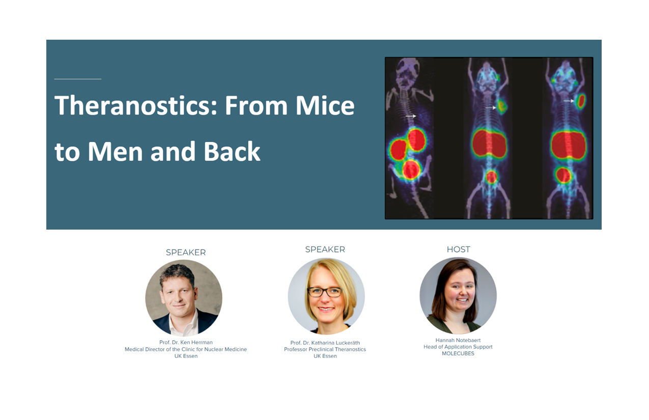
Theranostics: From Mice to Men and Back

Jun 25, 2024

The theranostic approach combines targeted therapy and diagnostic imaging. It represents a precision medicine approach relying on a specific targeted diagnostic test that helps to select patients for a specific targeted therapy. Molecular imaging in nuclear medicine combines imaging modalities like PET and SPECT with computed tomography (CT) or magnet resonance tomography to derive detailed information on disease. In the clinic, molecular imaging is mainly used for diagnosis, staging, monitoring response to therapy, and selecting patients. Diagnostic imaging can identify those patients who are positive or negative for a target out of a larger population. Subsequently, the patients who are positive can continue with targeted therapy, and patients who are negative can receive conventional therapy. Using preclinical imaging we can learn about targets, (the choice of) ligand, (potentially) which radioisotope to use best, and about tumor biology, which is important for effective theranostics.


Watch webinar after proceeding to registration

Scientific paper
Application

Related technologies: PET, SPECT, CT

Get more info

Irina Cojocaru

Sales manager

Irina

Cojocaru

+40 749 952 785

Send Message

Brand profile

MOLECUBES

PET/SPECT/CT preclinical imaging CUBES

Related products

The high-throughput CT preclinical imager

show detail

The high-performance PET preclinical imager

show detail

The high-sensitivity, high-resolution SPECT preclinical imager

show detail

We supply and support Life-Science Technologies in the territory of Central and Eastern Europe.

Czech Republic

Slovakia

Hungary

Poland

Croatia

Slovenia

Serbia

Romania

Bulgaria

Latvia

Lithuania

Estonia

Russia

Ukraine

Belarus

Turkey