
With Benchtop Microfluidic Cell Sorting, NanoCellect is committed to empowering every scientist to make discoveries one cell at a time.
Accelerate to discover
Back to filter
Related topics

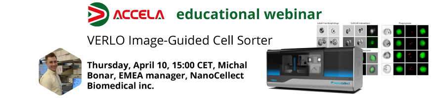
Webinar recording: Introducing VERLO Image-Guided Cell Sorter
Watch our webinar recording to learn more about NanoCellect VERLO Image Guided Cell sorter. VERLO is providing conventional flow cytometry and cell sorting along with the ability to perform imaging analysis to identify and sort cells based on morphology, subcellular localization and more. The VERLO instrument expands the capabilities of gentle benchtop microfluidic cell sorting. With two lasers and nine colors, plus 3 label-free parameters, it maintains simple workflows for either bulk sorting or single-cell dispensing into 96- or 384-well plates with an integrated cell dispensing and sample cooling.
- Spatial Information
Captures images of each cell. In addition to fluorescence intensity, the VERLO provides spatial information such as cell morphology and marker localization
- Enhanced Resolution
Imaging of subcellular structures and analysis of spatial variations in brightfield, darkfield, and fluorescent images
- Increased Precision Sorting
Following the acquisition and processing of cell images, imaging features are incorporated into the gating strategy to define cell phenotypes
- Multivariate Datasets
Combines high-throughput sampling with single-cell image acquisition and measures large number of features from cell images, providing more comprehensive data
In cooperation with Michal Bonar, EMEA manager, NanoCellect Biomedical inc.
Related technologies: Cell Sorting
Related products

BioFocus newsletter
Register to receive Brand new technology development, Important product updates, Interesting scientific events and more!
Technologies