Products
“I am very pleased to have the opportunity to work with the Lumascope, a compact microscope with very powerful autofocus that can fit in standard cell culture incubator, easily accessible for objective exchange. Fluorescent measurement showed high quality of pictures taken with crisp fluorescent signal, enabling to distinguish GFP labeled mitochondria using 40x lens. High definition phase contrast module enable also label free cell proliferation. Thus, the system is definitely a great asset to test the effect of compounds on proliferation and morphology changes”.
BIOCEV, Vestec, Czech Republic

“I am very pleased to work with a Lumascope inside the cell incubator which allow us to follow the cell proliferation and migration for days. More importantly, it is a versatile instrument to follow the apoptosis of fluorescently-labelled cells (e.g., CFSE) upon modulating the primary NK cell activity with various compounds in co-culture set-ups. The fluorescent images are of high resolution and quality over the investigated time frames. The microscope could also be used for following the formation of organoid structures in 3D experiments. This system is indeed a good device for live cell imaging while multiple conditions and compound are simultaneously screened”.
Grigore T. Popa University of Medicine and Pharmacy of Iasi, Romania Faculty of Medicine, Department of Immunology

“We have chosen Etaluma LS620 for its superb combination of optics, image quality, sturdiness and ability to operate at very high relative humidity inside an incubator. We are using it for cell culture under flow in microfluidic chips in tightly controlled environment using programmatic hypoxia modeling and other cell environment modeling conditions. Since LS620 is a digital inverted fluorescent microscope, it allows for parameter setup and image capture from distance reducing incubator environment interreference.”
Institute of Biology and Immunology of Reproduction, Sofia, Bulgaria

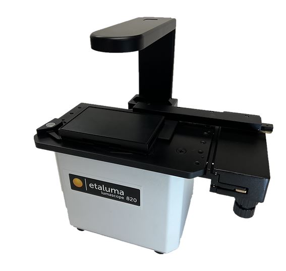
“The BioFlux 200 system in combination with Lumascope 720 microscope, help our research by visualizing cell interactions and monocyte to endothelium adhesion mechanisms under shear flow.The bundled solution proves a novel and attractive experimental approach, compared to conventional static live-cell assays.”
Uniwersytet Jagielloński (Collegium Medicum), Krakow, Poland

“I had a chance to test Lumascope in our cell culture lab. The device gives a great opportunity to observe live cells in a phase-contrast and fluorescence. We performed co-culture experiments where each cell type was labeled with different fluorochrome. Lumascope allowed for distinguishing cell type and observation of interaction between cells. The device was placed in a cell culture incubator with O2 control, therefore we could analyze those interactions in normoxic and hypoxic conditions. Additionally, the system can be useful for analysis of cells which express fluorochromes under control of certain promoters. It enables the evaluation of exact moment of certain genes activation and measurement of fluorescence intensity”.
Medical University of Warsaw, Warsaw, Poland

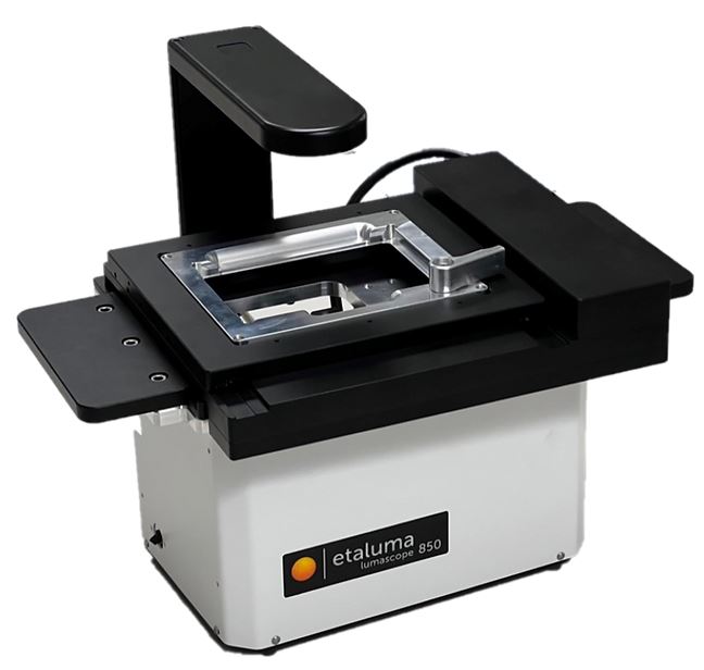
Lumascope 850 – Etaluma
Latest generation, 3 colors, fully-automated microscope
The powerful, new LS850 Microscope is the latest generation of our fully automated three-channel flagship model and offers the latest advances in optics, cameras, throughput, and user flexibility. . The LS850 improves the image quality, motion speed, illumination, and software flexibility over the previous LS720 model.
Exquisite XY motion control, motorized focus that allows autofocus and z-stacks, and easy-to-configure software combine to facilitate your automated microscopy experiments.
The LS850 in your incubator allows you to have a live cell imaging system that offers minimum photo toxicity and the most stable environment for long term imaging. Whether imaging multiple fields in your flasks or 1536 wells of cells with 3 fluorophores in a multi-day time-lapse, the LS850 offers a whole new world of walk-away automated microscopy.
Key Features:
- Images, tiled images, Z-stacks, time-lapse series, and videos
- Bright field and phase contrast transmitted illumination
- Multi-OS software allows set-up and control across any locations, including microplates, microfluidic chips, slides, dishes, flasks, and deck-top chambers and custom arrays
- Compact and robust design enables use inside cell culture hoods, incubators, hypoxic chambers, and gloveboxes
- Detects blue, green and red fluorophores, including common probes such as Hoechst, DAPI, FITC, Fluo-4, GFP, Texas Red & mCherry GFP
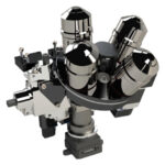
- Objective compatibility with standard lenses and automated 4-objective turret
More info at: https://etaluma.com/products/lumascope-850/
Setup a demonstration
with our specialist

