Products

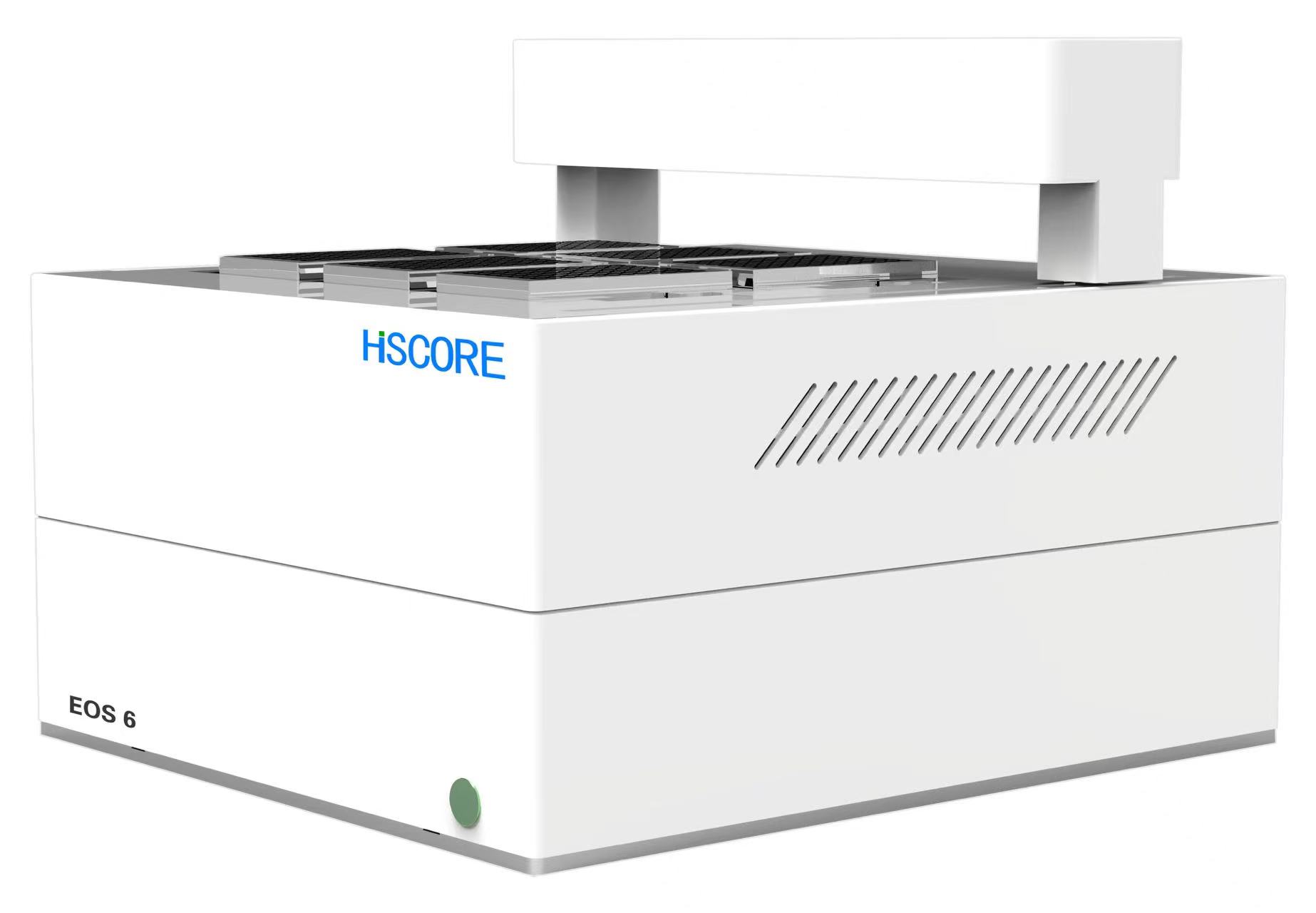
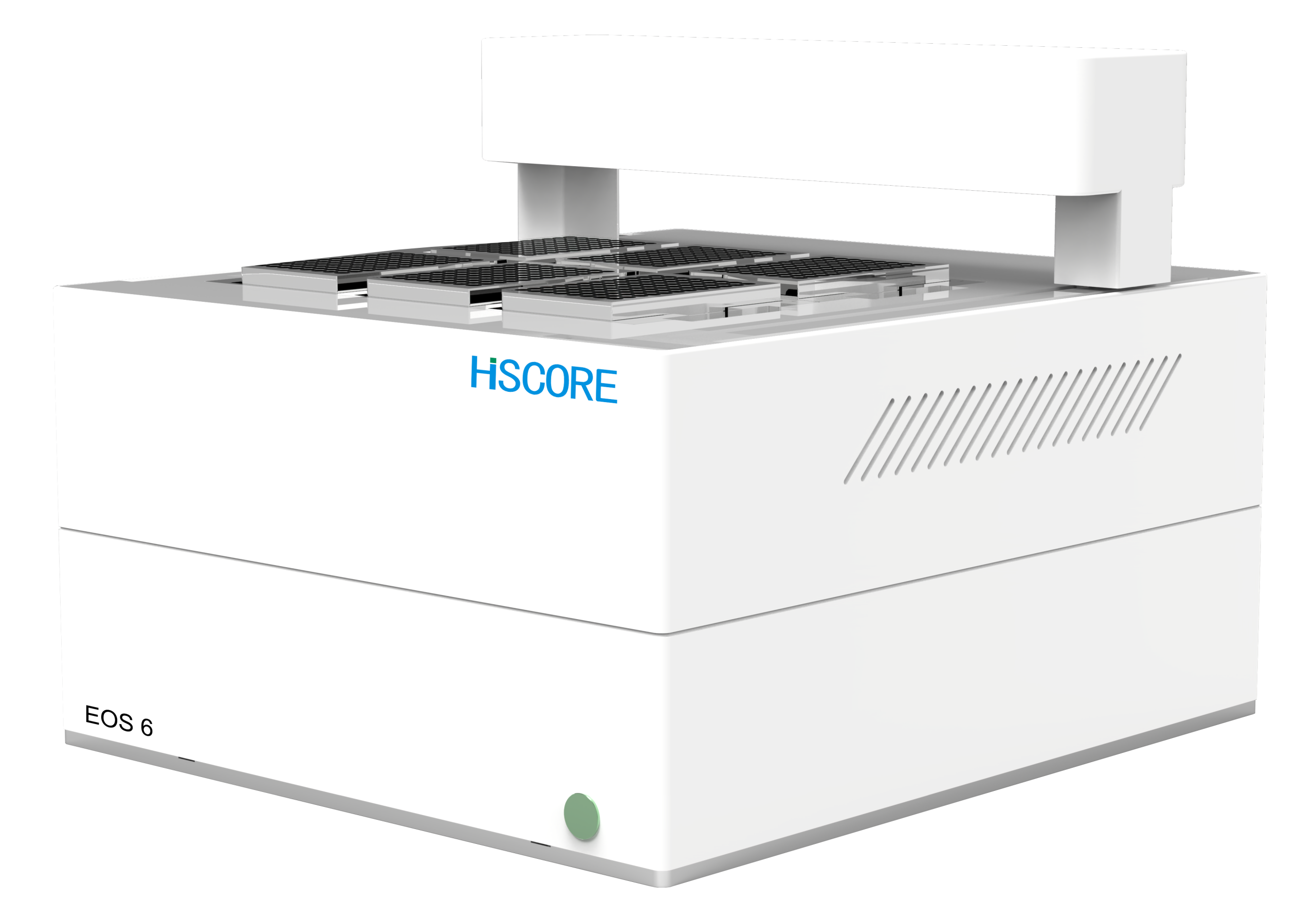
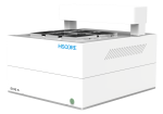
EOS 6 live-cell analysis system
High-throughput live cell analysis inside your incubator
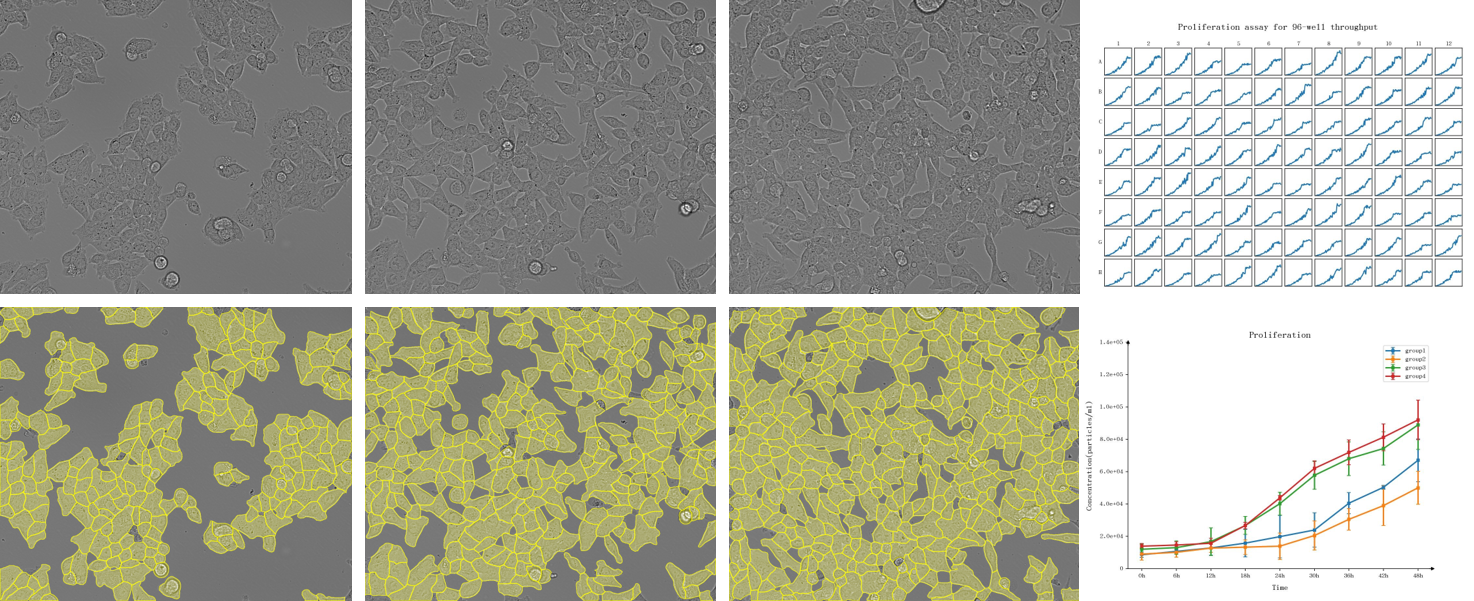
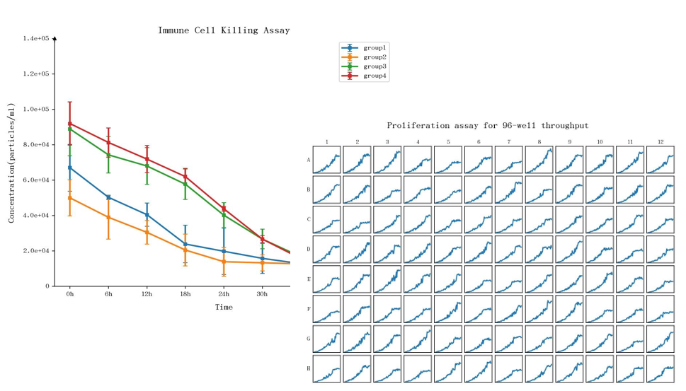
Eos6 Live-Cell Analysis System is designed to efficiently capture cellular changes where they happen – in the incubator. Capture high-resolution images and record data in real time over hours, days or weeks. From proliferation assays to immune killing of tumor spheroids, this flexible system enables users to observe and quantify complex biological changes. Integrated software simplifies data analysis to speed time to answer while producing publication-quality graphs and plots.
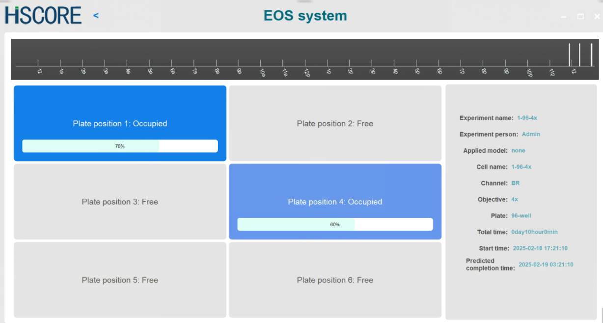
The “power of 6” all-in-one platform
Built to support multiple users and applications, the EOS6 can run up to six microplates in parallel, flask, dishes, slides… Users can schedule experiments at different image acquisition frequencies and magnifications in parallel.
Optical system:
Optimized for Olympus objective lens on an automated turret (4X, 10X, 20X, 40X) , ensuring high quality images and experimental flexibility from whole well imaging to intracellullar observation. The optical system is designed to withstand the temperature and humidity conditions of a standard cell culture incubator, allowing the instrument to be placed inside a CO₂ incubator for long-term time-lapse imaging.
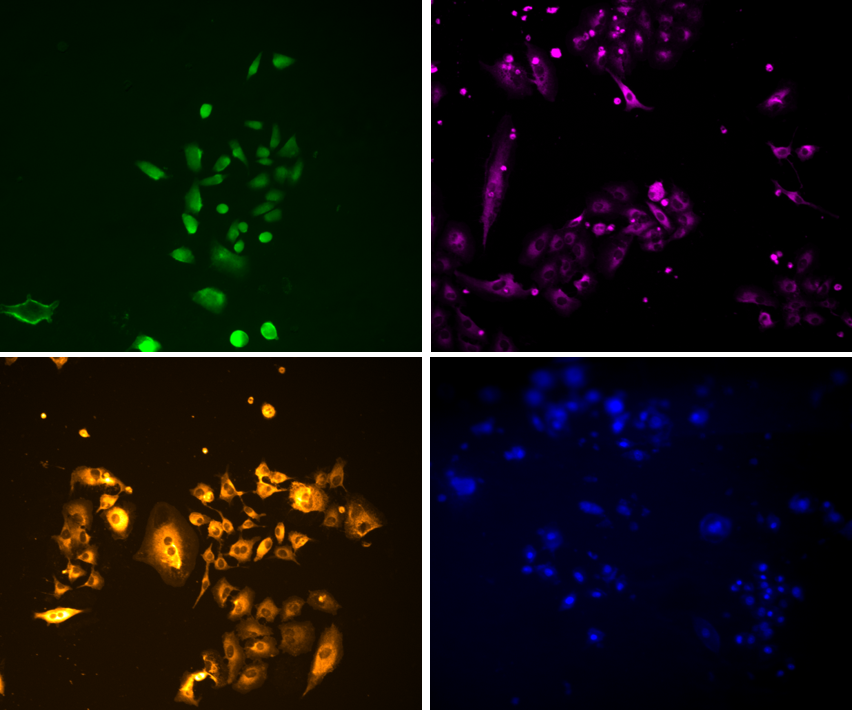
Imaging mode:
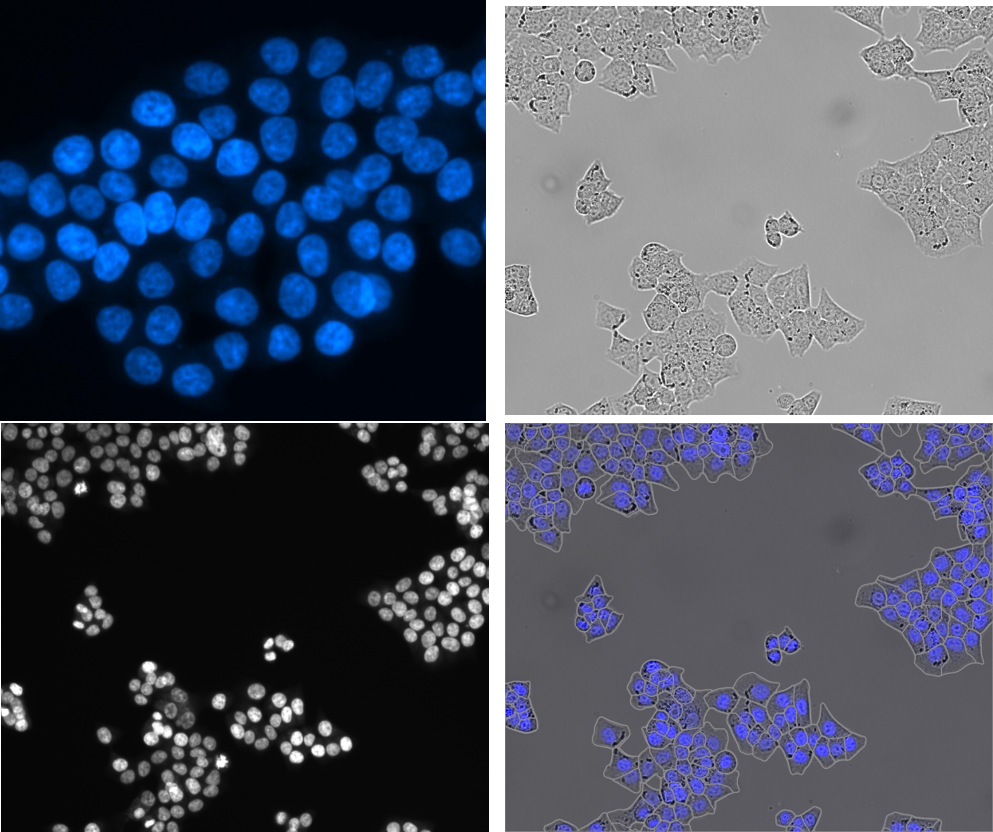
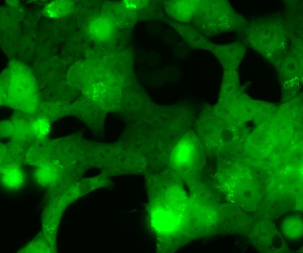
Bright field, phase contrast, monochromatic fluorescence, 4 fluorescence channels (red, green, blue and NIR), Z-Stacking, whole-well imaging.
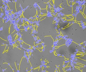
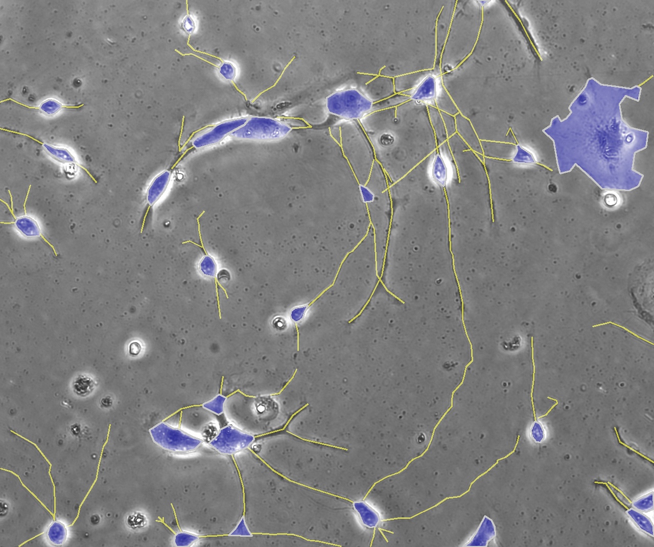
AI powered application modules for demanding applications:
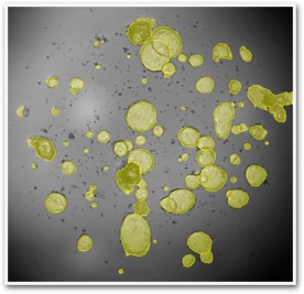
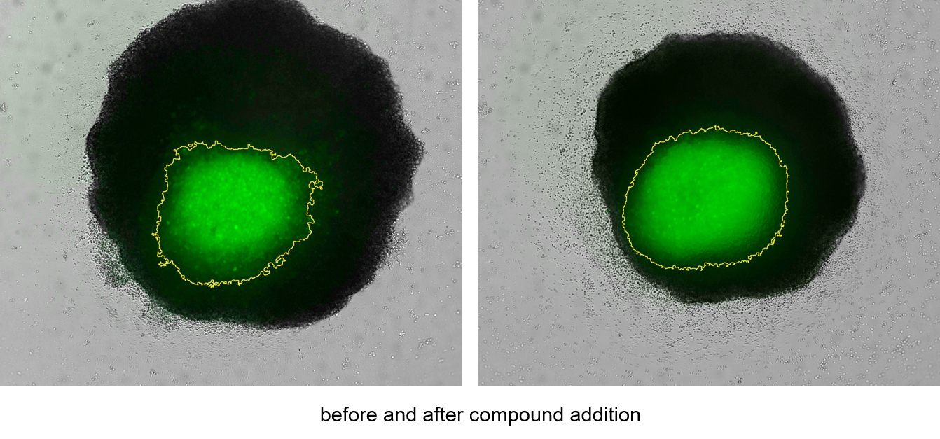
Intuitive software interface combined with AI-powered algorithms enables more accurate cellular segmentation and analysis. Application modules are specifically developed to support demanding experimental workflows as : Organoids, Label free Cell count, 3D Spheroids and Organoids, Cell migration/Invasion, Chemotaxis, Immune cell killing, Phagocytosis, Angiogenesis and Neuronal Cell proliferation.
Documentation

Setup a demonstration
with our specialist
Related products


BioFocus newsletter
Register to receive Brand new technology development, Important product updates, Interesting scientific events and more!
Technologies